Episode 4: Ovarian Torsion
Discover the 4 D’s of Radiology DETECT - DESCRIBE - DIFFERENTIAL - DECISION
DETECT
Epidemiology
- Occurs in 2-3% of patients that present with pelvic pain
- Preferred terminology: adnexal torsion as the fallopian tube is often involved
- Risk Factors:
- Benign ovarian lesion causing enlargement (especially masses > 5 cm ): Cysts, Cystic Teratomas
- Fertility treatment such as ovulation induction
- Pregnancy
- Previous pelvic surgery
- Age: Most commonly women of child bearing age
Anatomy
Understanding the anatomy is key to grasping the pathophysiology of which structures twist to result in vascular compromise
Ovarian Ligaments:
- Broad Ligament: Attaches the uterus to the pelvic side wall. Includes the mesovarium, mesometrium and mesosalpinx
- Suspensory Ligament of ovary: Often thought to be a part of the broad ligament, attaching the ovary to the pelvic side wall and houses the ovarian artery, vein, nerves and lymphatic tissue
- Ovarian Ligament: Ovary to Uterus
Ovarian Blood Supply and Venous Drainage
- Arterial: Dual blood supply from the ovarian artery and adnexal branches of the uterine artery. This is important as it explains why arterial compromise,is often a late finding
- Venous: Ovarian vein and uterine veins. The right ovarian vein drains directly into the IVC where as the left drains into the left renal vein
Pathophysiology
The ovary and fallopian tube twist on the suspensory ligament. The right ovary is more commonly involved often secondary to the mobility of the ileum and cecum as compared to the rectosigmoid colon
Perfusion is disrupted leading to venous congestion, lymphatic congestion and then compromised arterial flow
Outcomes if left untreated include ovarian infarction and subsequently subfertility
Clinical Presentation
- Acute abdomen: Sudden and severe abdominal pain that often warrants surgical referral
- Pain: Can be intermittent due to twisting and untwisting of the vascular pedicle
- Nausea and Vomiting
- Less common: Fever, palpable abdominal mass
DESCRIBE
Imaging Workup
First line imaging modality is ultrasound (Transabdominal, Transvaginal + Doppler) when clinically suspecting ovarian torsion or other gynecological causes of acute pain
CT +/- Contrast may be considered based on clinical presentation
MRI in certain clinical circumstances and when available
Ultrasound Findings
Findings:
- Ovarian Findings
- Enlarged ovary > 4cm
- Displacement of the ovary midline
- Increased central echogenicity of the ovary
- Peripheral follicles (look like avascular hypoechoic cystic structures). As the center of the ovary increases in size due to edema, the follicles located within the cortex are pushed to the side leading to this “string of pearls” quality
- Follicular ring sign: Hyperechoic rim surrounding the displaced follicles
- Doppler Assessment
- Abnormal arterial flow is often a late finding
- Venous flow abnormalities including non-continuous or absent flow occur prior to arterial abnormalities. Highly sensitive and specific for ovarian torsion
- Blood supply may be normal due to twisting and untwisting as well as the dual blood supply of the adnexa
- Tube/Ligamentous Suppor
- Twisted whirlpool or target sign: Often subtle finding
- Abnormal blood flow
- Peritoneum
- Free fluid
Previous Imaging
- Ovarian cyst
- Cystic Teratoma: Cyst with hyperechoic lesions from sebaceous or fatty material with internal markedly hyperechoic calcifications and associated posterior acoustic shadowing
The benefits of ultrasound is the real time nature and ability to ask the pertinent history questions that will fuel your differential
CT Findings
High negative predictive value if both ovaries are visualized. Can be helpful in the context of an indeterminate ultrasound
Findings
- Ovary:
- Ovarian enlargement
- Location: often displaced midline
- Lead mass with smooth regular wall thickening. Presence of irregular wall thickening is suggestive of nonviability
- Lack of ovarian enhancement is a later stage finding suggestive of nonviability
- Tube and Pedicle
- Twisted pedicle whirlpool
- Triangular enhancing soft tissue thickening on the lateral aspects of the uterus. Can be difficult to differentiate this as a separate entity from an adnexal mass
- Distended vessels can be visualized on contrast-enhanced imaging
- Uterus
- Displaced towards the side of the involved ovary
- Peritoneum
- Fat stranding and free fluid surrounding involved adnexa
- Hemorrhagic Infarction: Predictive of ovarian nonviability and necrosis
DIFFERENTIAL
DDX
The history is your guide
ACUTE ABDOMINOPELVIC PAIN
Gynecological
- Ruptured hemorrhagic ovarian cyst/Hemorrhagic corpus luteal cyst
- Findings:
- Internal low level echoes with a dependent portion
- Internal echoes organize into a fibrinous web – thin and avascular and do not extend across entire cavity
- Retracted clot: Heterogenous avascular mass with outward concavity. Apply pressure and it may change shape
- Free fluid within the peritoneal cavity
- Thin rim of non-edematous ovarian tissue
- Ovarian Hyperstimulation
- Cause: Fertility treatments involving ovarian stimulation
- Clinical Presentation: Abdominal or pelvic pain with bloating
- Findings
- Ovarian Findings: Enlarged follicles around periphery of ovary with normal central ovarian tissue. Often bilateral
- Associated findings: Ascites, pleural effusions
- These can be at risk for torsion
- Ectopic pregnancy
- Please refer to our previous podcast
- Endometriosis with endometriomas
- Findings of an endometrioma:
- Cystic structure with internal homogenous low level echoes
- No internal flow
- Leiomyoma degeneration
- What: pedunculated leiomyomas that undergo degeneration
- Challenge: May be difficult to differentiate on CT
- Malignant Masses
- Tubo-Ovarian Abscess
- Findings
- Complex solid/cystic adnexal mass
- Thick walled structure with internal septations and debris
- Location: Can be bilateral
- Associations: Pyosalpinx appearing as a dilated adnexa +/- internal echoes
Non-Gynecological
- Renal Pathology: Ureteric calculi, cystitis
- Bowel athology: Inflammatory Bowel disease, Diverticulitis, Appendicitis
DECISION
Game Plan
- Appropriate gynecology referral
- Pain Management
- Surgical exploration and treatment
- Maintenance of fertility is the mainstay of care
- Laparoscopic exploration may be utilized to assess ovarian viability
- Detorsion often occurs +/- cystectomy even in the case of an ovary that looks blue/black
- Cystectomy may be delayed preventing further insult to the affected ovary
- Oophorectomy is considered in patients presenting with high risk of malignancy or in post-menopausal patients
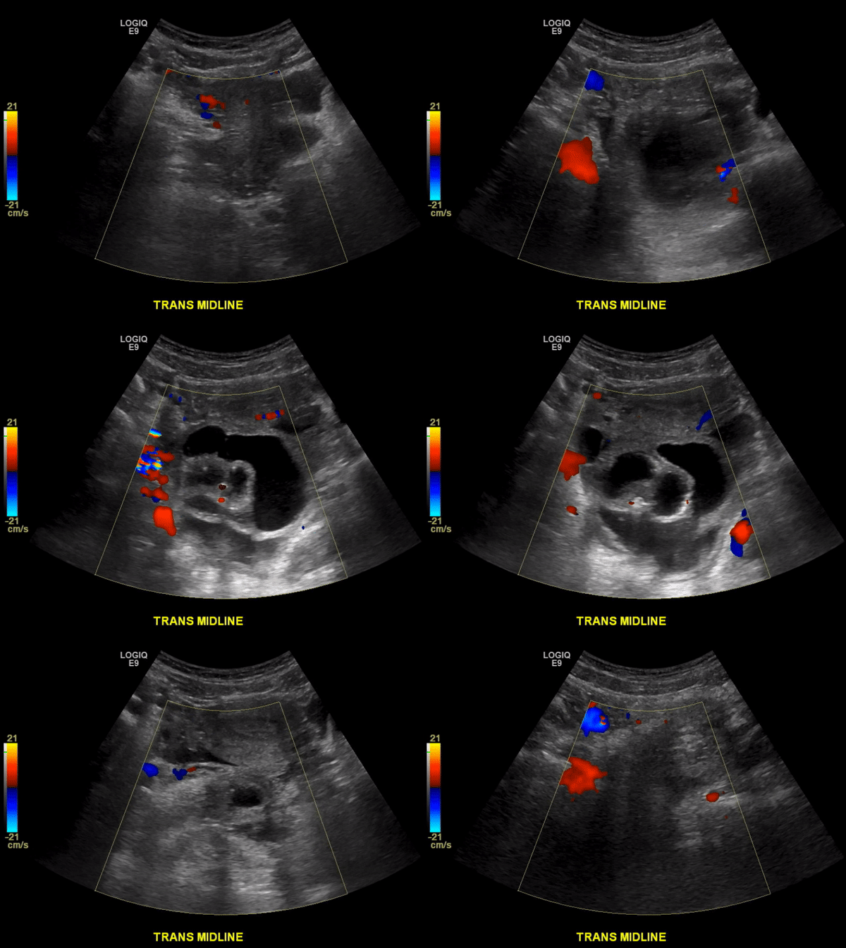
Example Case of Ovarian Torsion
References
Dawood, M. T., Naik, M., Bharwani, N., Sudderuddin, S. A., Rockall, A. G., & Stewart, V. R. (2021). Adnexal Torsion: Review of Radiologic Appearances. RadioGraphics, 41(2), 609-624.
Chang, H. C., Bhatt, S., & Dogra, V. S. (2008). Pearls and pitfalls in diagnosis of ovarian torsion. Radiographics, 28(5), 1355-1368.
Mashiach, R., Melamed, N., Gilad, N., Ben-Shitrit, G., & Meizner, I. (2011). Sonographic diagnosis of ovarian torsion: accuracy and predictive factors. Journal of Ultrasound in Medicine, 30(9), 1205-1210.
Nalaboff, K. M., Pellerito, J. S., & Ben-Levi, E. (2001). Imaging the endometrium: disease and normal variants. Radiographics, 21(6), 1409-1424.
Chavhan, G. B., Parra, D. A., Mann, A., & Navarro, O. M. (2008). Normal Doppler spectral waveforms of major pediatric vessels: specific patterns. Radiographics, 28(3), 691-706.
Kives, S., Gascon, S., Dubuc, É., & Van Eyk, N. (2017). No. 341-diagnosis and management of adnexal torsion in children, adolescents, and adults. Journal of Obstetrics and Gynaecology Canada, 39(2), 82-90.
Munro, K., Gharaibeh, A., Nagabushanam, S., & Martin, C. (2018). Diagnosis and management of tubo‐ovarian abscesses. The Obstetrician & Gynaecologist, 20(1), 11-19.
